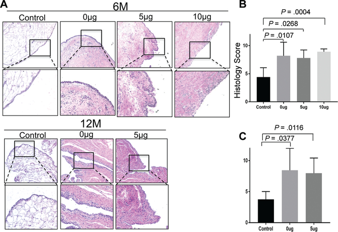
Figure 5.
Microscopic evaluation of synovitis. (A) Representative photomicrographs of synovium stained with hematoxylin and eosin at 6 and 12 months. Histology score at (B) 6 months and (C) 12 months (0, best; 12, worst). The data are presented as mean ± SD. P value was calculated with the Kruskal-Wallis test, followed by post hoc Dunn multiple-comparison tests.

