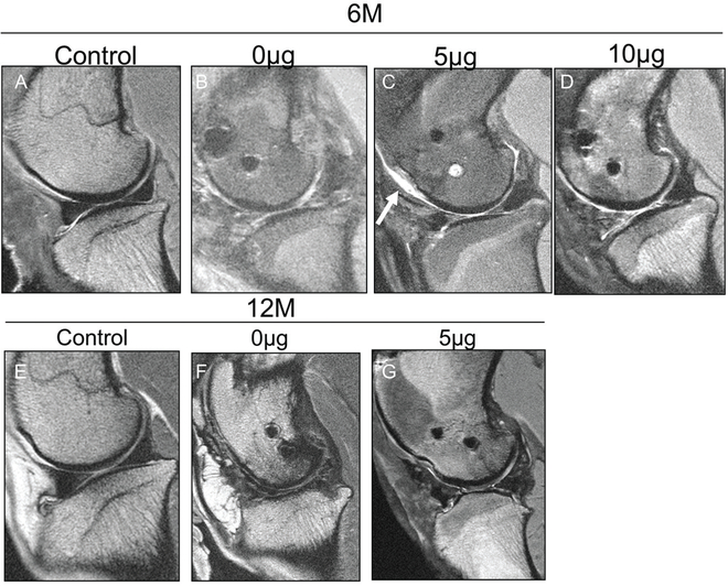
Figure 7.
Magnetic resonance imaging evaluation of articular cartilage of medial compartment. Representative sagittal 2-dimensional fast spin echo images of the sheep knee with a meniscal construct implanted in the medial compartment evaluated at 6 and 12 months postoperatively. (A) Control specimen at 6 months displays preservation of the articular cartilage with a hypointense meniscus and no extrusion present. (B) Specimen with 0-μg meniscal scaffold at 6 months displays marked hyperintensity of the meniscal scaffold with extrusion of the anterior horn relative to the tibia, encased in thickened synovium. (C) Specimen with 5-μg meniscal scaffold at 6 months displays truncation of the free edge of the anterior and posterior horns, but the meniscus is not extruded and there is relative preservation of the hypointense signal. Chondral wear appears more severe over the trochlea (arrow). (D) Specimen with 10-μg scaffold at 6 months demonstrates marked hyperintensity in the anterior horn but no extrusion and with superficial chondral wear. (E) Control specimen at 12 months displays preservation of the articular cartilage with no meniscal extrusion present. (F) Specimen with 0-μg scaffold at 12 months demonstrates areas of full-thickness loss of articular cartilage and hyperintensity of the meniscal construct but only minimal anterior extrusion. (G) Specimen with 5-μg scaffold at 12 months demonstrates moderate extrusion of the anterior horn with truncation of the free edge and preservation of articular cartilage over the condyle, but there is focal wear over the tibia.

