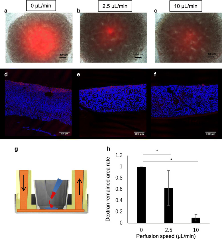
Fig. 5.
Dextran labeled with Texas Red dropped on the organoid cultivated for 2 days under static or perfusion conditions. a–c Microscopic merged images of Texas Red fluorescence and phase contrast images. Red color indicates the remaining Texas Red-labeled dextran. d–f Texas Red-labeled dextran and nuclear staining merged images. g Schematic explaining the dropped dextran at the start of cultivation. h Summary remaining dextran in the sections from (d–f) (n = 3)

