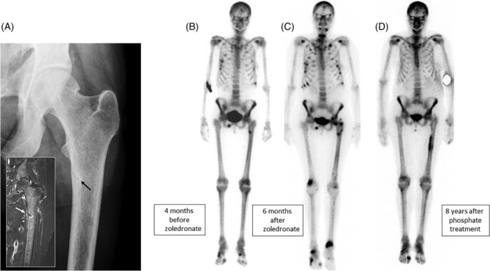
Figure 1.

Radiographic images from subject A. (A) Looser zone (pseudofracture) of the upper femur (inset T2‐STIR MR image) that occurred 4 months before zoledronate was administered. This was originally interpreted as a “stress fracture.” (B) Bone scintigram taken at the same time as the images shown in A. There is increased uptake of isotope at the site of the Looser zone, but also in several ribs and the right inferior pubic ramus. (C) Bone scintigram taken 6 months after the first zoledronate infusion. There are numerous new lesions in the ribs, both femora and the distal tibias. (D) Bone scintigram taken 8 years after phosphate treatment was started. There is increased uptake in relation to the left femoral rod and degenerative disease in the feet, but the other lesions have healed.
