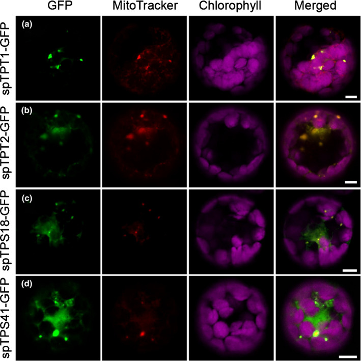
Figure 8.

Subcellular localisation of the four mitochondrial proteins. (a, b) Localisation of trans‐prenyltransferase (TPT) 1 and 2, respectively. (c, d) Localisation of terpene synthase (TPS) 18 and 41, respectively. Sequences corresponding to the putative mitochondria transit peptide (sp) were fused to a downstream GFP and transiently expressed in Arabidopsis leaf protoplasts. GFP fluorescence indicates the location of each fusion protein (shown in green), the location of chloroplasts was determined by chlorophyll autofluorescence (shown in magenta), and the localisation of mitochondria was determined by the fluorescence of MitoTracker (shown in red). Column labelled ‘Merged’ represents all the combined fluorescent signals. Bars, 5 μm.
