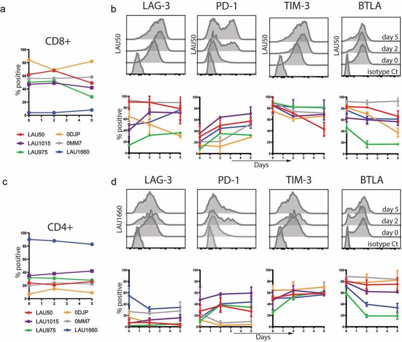
Figure 2.

Characterization of Inhibitory Receptors surface expression on TILs during co-culture with autologous melanoma cell lines. TILs from the indicated patients were stained with the indicated mAbs before co-culture (day 0) or 2 and 5 days after the co-culture with autologous melanoma cell lines. Percentages of CD8+ (a) and CD4+ (c) in the TILs are shown. Average with Standard Deviation of at least two independent experiments per patient and representative histograms of the indicated molecules for CD8+ (b) or CD4+ T cells (d) are shown.
