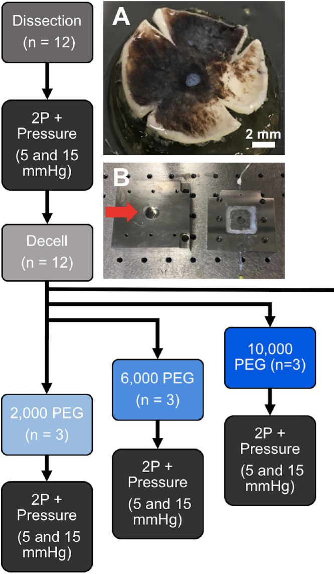
Figure 1.

Design of pressurization and LC displacement experiment. Dissected porcine posterior poles were cut into a clover-leaf pattern (A) and positioned into a specialized pressurization chamber (B), designed to allow for visualizing the LC using two-photon microscopy while controlling the pressure upon the tissue. The overall experimental design is illustrated in the flow chart. Scale bar = 2 mm.
