Figure 5.
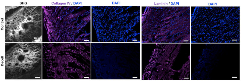
SHG and immunohistochemistry imaging of the decellularized posterior poles and non-decellularized controls. Histology was performed to compare the effect of decellularization on cellular content, ECM microstructure, and basal lamina of the tissue. SHG imaging of the control and decellularized posterior poles indicated that decellularization had minimal impact on ECM microstructure. DAPI (blue) staining validated the results from H&E staining, with the decellularized samples showing negligible cell nuclei compared to the controls. Tissues were stained for collagen IV (magenta) and laminin (magenta) to visualize the basal lamina, with similar immunofluorescence staining in the decellularized samples compared to the controls, indicating minimal damage to the basal lamina. Scale bar = 100 µm.

