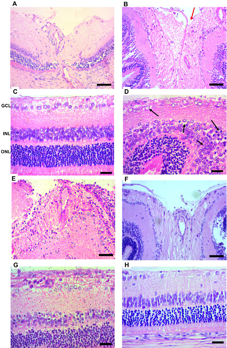
Figure 9.
Histologic analysis of healthy and ocular hypertension rats. The vehicle-treated ocular hypertension animals exhibited an increase in the excavation of the optic nerve (red arrows) and increase in the number of cytoplasmic vacuolization in the GCL (black arrows) (B, D) if compared to healthy rats (A,C). Besides, the INL displays more edema, pyknotic nuclei, and cellular disorganization (black arrows) (D). The ONL exhibits a decreased cell number, and greater edema and cell disorganization compared with the healthy retina D. Treatment with PnPP-19 (E,G) reduced histologic damage if compared to vehicle-treated glaucoma B and D or Bimatoprost (F,H). Healthy A and C; ocular hypertension vehicle-treated B and D; ocular hypertension treated with PnPP-19 E and G Bimatoprost F and H. Retina layers: ganglion cell layer (GCL), inner nuclear layer (INL), and outer nuclear layer (ONL). Digital images were obtained using a microscope (Apotome.2, Zeiss, Germany) with a 10× and 20× objective, scale bar = 50 µm.

