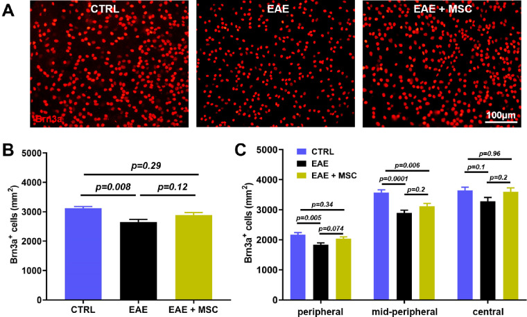
Figure 4.
MSC treatment alleviates RGC loss in EAE mice. (A) Representative images of Brn3a immunolabeling (red) display differences in RGC density in the peripheral retina of controls, EAE mice, and MSC-treated EAE mice. (B) Analysis of the average RGC density reveals a significant loss of Brn3a+ cells in untreated EAE mice and a remarkably higher RGC density in MSC-treated mice. (C) Regional differences in the RGC density between controls and EAE mice is most prominent in the mid-periphery. A notably upward trend in RGC numbers of EAE mice with MSC treatment is observed in the peripheral and central region, although this is not statistically significant.

