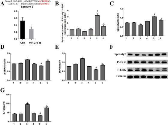
FIGURE 2.

A, DNA fragments within the 3′UTRs of the sprouty2 genes that contain the miR‐27a‐3p binding site were cloned into the luciferase reporter. Luciferase activity in the cells was measured. It confirmed that mir‐27a‐3p targets Sprouty2 gene. B and C, Real‐time PCR and Western blot were used to observe Sprouty2 mRNA and protein expression. D‐F, Western blot was used to observe pERK and tERK protein expression. G, IL‐10 was examined by ELIAS. Data from three independent experiments; mean ± SD. * P< 0.05 for the difference compared with control and mimic NC group; # P< 0.05 for the difference compared with pcDNA3.1 group. ^P< 0.05 for the difference compared with the pcDNA3.1‐sprouty2 group. 1, control group; 2, mimics NC group; 3, mir‐27a‐3p mimics group; 4, pcDNA3.1 group; 5, pcDNA3.1‐sprouty2 group; 6, mir‐27a‐3p mimics‐pcDNA3.1‐sprouty2 group
