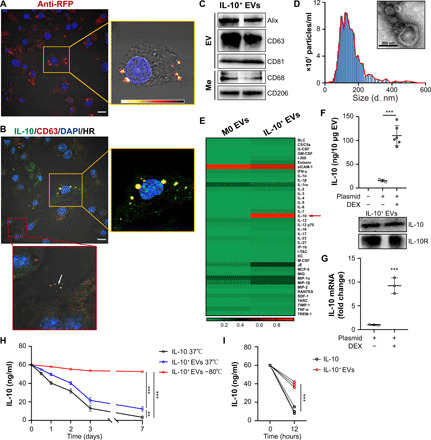
Fig. 1. Preparation and characterization of the IL-10+ EVs.

(A) RAW cells were transfected with RFP-tagged IL-10 and then were stimulated with dexamethasone. The intracellular distribution of IL-10 was examined by immunostaining against RFP. The right box is a heatmap visualization of the boxed region. Scale bar, 10 μm. (B) Immunostaining of IL-10 (green) and the EV marker CD63 (red) in engineered RAW cells. The yellow box is a higher magnification of the boxed region in the merged image. Scale bar, 10 μm. (C) Western blotting analysis of EV-associated (Alix, CD63, and CD81) and macrophage-associated markers (CD68 and CD206) in IL-10+ EVs. (D) Size distribution and representative TEM images of IL-10+ EVs. (E) A heatmap showing the protein levels (normalized to array reference) of each cytokine obtained from antibody array analysis. M0 EVs, EVs from untreated RAW cells. G-CSF, granulocyte colony-stimulating factor; GM-CSF, granulocyte-macrophage colony-stimulating factor; IFN-γ, interferon-γ; M-CSF, macrophage colony-stimulating factor; TNF-α, tumor necrosis factor–α. (F) ELISA analysis of IL-10 in EVs, and Western blotting analysis of IL-10 and IL-10 receptor in IL-10+ EVs (n = 3 or 6). (G) IL-10 mRNA in IL-10+ EVs was measured by real-time quantitative PCR (n = 3). (H and I) Comparison of the stability of IL-10+ EVs and free IL-10 under different conditions, including put in 37° or − 80°C for a week, suspended in pH 5.5 solution for 12 hours. Then, IL-10 concentration was detected by ELISA analysis at different time points (n = 3). Data are presented as means ± SD. **P < 0.01 and ***P < 0.001, two-tailed t test (F, G, and I) and one-way analysis of variance (ANOVA) (H). BLC, B lymphocyte chemoattractant; IP-10, interferon gamma induced protein 10; I-TAC, interferon-inducible T-cell alpha chemoattractant; KC, C-X-C motif chemokine 1; JE, monocyte chemoattractant protein-1; MCP-5, monocyte chemoattractant protein-5; MIG, monokine induced by interferon-gamma; MIP-1a, macrophage inflammatory protein 1α; RANTES, regulated on activation, T-cell expressed, and secreted; SDF-1, stromal cell-derived factor 1; TARC, thymus and activation regulated chemokine; TIMP-1, tissue inhibitor of metalloproteinases 1; TREM-1, triggering receptor expressed on monocytes 1.
