
Fig. 4. Molecular and cellular deficits in Vamp2rlss.
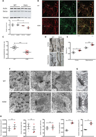
(A) Western blots of whole-brain lysates in WT (gray) and homozygotes (Vamp2rlss, red). STX1a and VAMP2 are quantitated relative to actin levels. (B) WT and Vamp2rlss hippocampal cell cultures immunostained with VAMP2 (red) and BASSOON (green); colocalization is shown in merged channel images (yellow, right panels). Arrows indicate presynaptic active zone containing both VAMP2 and BASSOON. Insert shows a close up of stained neuron showing VAMP2, BASSOON, and colocalization. Scale bars as shown. (C) VAMP2 expression in DIV15 primary cell culture. VAMP2 fluorescence intensity was normalized to that of BASSOON. (D) Golgi-stained sections showing mushroom (white arrowhead), long (black arrowhead), and stubby (white arrow) spines. Scale bar, 10 μm. (E) Spine counts per unit length in WT (gray) and Vamp2rlss (red) samples. (F) Electron micrographs of hippocampal sections. Scale bars as shown. (G) Close-up of images in (F) showing docked vesicles (arrows). Scale bars as shown. (H) Measurement of synaptic parameters in WT (gray) and Vamp2rlss (red) mice. Individual data points are shown as is mean ± SEM, *P < 0.05, **P < 0.01, ***P < 0.001.