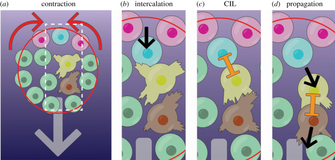
Figure 6.
Stages of supracellular neural crest cell migration. (a) Cells at the edge of the cluster are linked by an actomyosin cable (red). The cable contracts at the rear (red cable, red arrows indicate contraction) but not at the front. (b) All cells at the rear are brought closer together, causing cells to intercalate forwards (blue cells intercalation as pink cells move closer together; movement is black arrow). (c) The intercalating cell (blue) makes contact with the unpolarized cell in front (yellow), causing it to polarize, producing protrusions forward. This occurs by CIL (orange inhibition symbol). (d) This cell (yellow) then moves forward, propagating the signal to the cells in front (brown cell), and so forth. Thus, an anterograde wave of aligned forward cell flow emanates from the rear of the cell cluster.

