Figure 6.
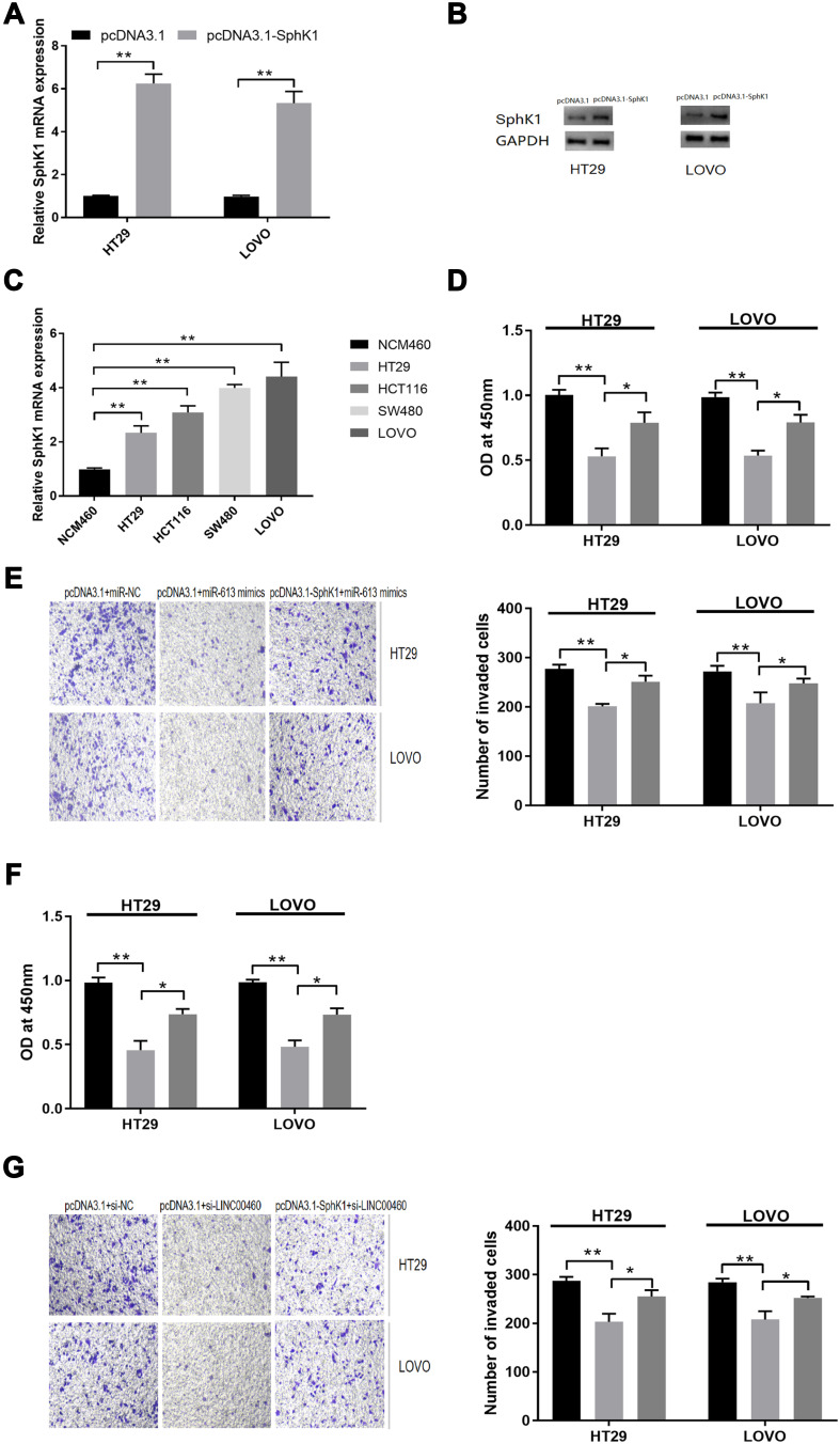
SphK1 is targeted by miR-613 in HT29 and LOVO cells. (A) qRT-PCR analysis and (B) Western blot analysis of SphK1 mRNA and protein expression levels in HT29 and LOVO cells after pcDNA3.1 or pcDNA3.1-SphK1 transfection. (C) qRT-PCR analysis of SphK1 mRNA expression levels in normal cell line (NCM460) and CRC cell lines (HT29, HCT116, SW480 and LOVO). (D) Cell proliferation and (E) cell invasion of HT29 and LOVO cells co-transfected with pcDNA3.1+miR-NC, pcDNA3.1+miR-613 mimics, or pcDNA3.1-SphK1+miR-613 mimics were detected by CCK-8 assay and Transwell invasion assay, respectively. (F) Cell proliferation and (G) cell invasion of HT29 and LOVO cells co-transfected with si-NC + pcDNA3.1, si-LINC00460+pcDNA3.1, si-LINC00460+pcDNA3.1-SphK1 were detected by CCK-8 assay and Transwell invasion assay, respectively. *p < 0.05, **p < 0.01.

