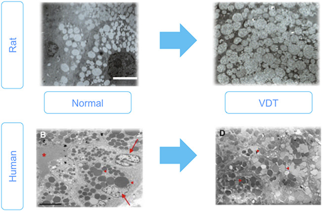
FIG. 8.

Lacrimal gland hypofunction in visual display terminal (VDT) users and animal model. Upper Figures: Rat VDT user model causes alterations in lacrimal gland morphology. Electron microscopic analysis of acinar cells of normal and VDT rat model. Images showing expanded acinar cells accompanied by accumulated enlarged secretory vesicle in the cytoplasm, decreased endoplasmic reticulum. Scale bar = 10 micrometers. Lower Figures: Electron microscopic findings of the lacrimal gland acinus in human normal and VDT user. Homogeneous secretory vesicles in normal controls. Scale bar = 5 micrometers. High magnification view of secretory vesicles in the VDT user. Red arrows, arrowheads and asterisk show nuclei, secretory vesicles and ductal lumen, respectively. Figures from77,79 Nakamura et al. and Kamoi et al., respectively. These are open access articles distributed under the terms of the Creative Commons Attribution License.
