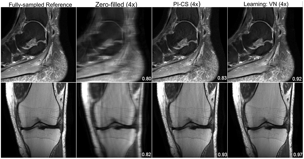
Figure 3.

From left to right: A single slice of the reference, zero-filled, PI-CS and VN reconstructions of a sagittal, proton-density weighted, fat-suppressed ankle image (top) and a coronal proton-density weighted knee image (bottom). The displayed structural similarity index (SSIM) was calculated for the presented slice. The VN reconstruction has less noise amplification and residual artifact than the PI-CS reconstruction. The sequence parameters were as follows: ankle – sagittal fat-saturated proton-density (PD-FS): TR = 2800 ms, TE = 30 ms, turbo factor (TF) = 5, matrix size = 384 x 384, in-plane resolution 0.42 x 0.42 mm2, slice thickness = 3.0 mm; knee – coronal PD: TR = 2750 ms, TE = 32 ms, TF = 4, matrix size = 320 x 320, in-plane resolution = 0.44 x 0.44 mm2, slice thickness = 3.0 mm.
