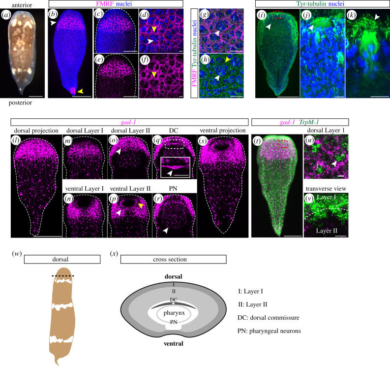
Figure 2.
Neurons are present in high density in the anterior and are organized into discrete structures including putative sensory organs. (a) H. miamia juvenile animal, dorsal view. (b) FMRF immunostaining concentrated in the anterior (white arrowhead). A small number of cells in the tail tip are also labelled with this marker (yellow arrowhead). (c,d) Zoom-in of anterior showing reticulated neurite bundles with (c) and without (d) nuclei. (e,f) Further zoom-in with (e) and without (f) nuclei showing clusters of nuclei (white arrowhead) surrounded by a reticulated network of neurite bundles (yellow arrowheads). (g,h) Anti-Tyr-tubulin (green) labels neurite bundles (yellow arrowhead) labelled by anti-FMRF and cells at the centre of the nuclei clusters (white arrowheads). (i) Tyr-tubulin labels frontal organ (dashed red box; shown in higher magnification in (j)) and putative sensory cells (white arrowhead; shown in higher magnification in (k)). (j) a high magnification view of the frontal organ (white arrowhead). (k) a high magnification view of a putative sensory cell with cilia (white arrowhead). (l–s) gad-1 mRNA expression showing layering in the anterior condensation and major neural structures. (l) Dorsal projection with gad-1+ cells concentrated in the anterior and diffusely distributed in the posterior. (m,n) Layer I contains a neurite bundle network. (o,p) Layer II contains mesenchyme-like cell bodies (white arrowheads). (p) Oral nerve ring located ventrally (yellow arrowhead). (q) Dorsal commissure (DC; white arrowhead) located above pharynx. Dashed white box outlines region highlighted in high magnification inset (solid white box). (r) Pharyngeal neurons (PN; white arrowhead). (s) Ventral projection with gad-1+ cells recapitulating the pattern seen in the dorsal view. (t) TrpM-1 is expressed in clusters of cells throughout the body, concentrated in the anterior (dashed red box; shown in higher magnification in (u)). (u) TrpM-1 expression in groups of clustered cells (white arrowhead) that correspond to nuclei clusters surrounded by gad-1+ neurite bundles within Layer I. (v) Transverse view through Layer I and Layer II (boundary between layers is denoted by dashed white line) showing that the TrpM-1+ cells are contiguous between the two layers. (w) A schematic of H. miamia showing location (dashed black line) of cross section through the anterior condensation shown in (x). (x) Schematic of cross section through the anterior condensation summarizing the neural anatomy revealed by the gad-1 mRNA expression. Dashed white lines around specimens show the outline of the animal. Scale bars: (a,b) 200 µm, (c,d) 100 µm, (e,f) 10 µm, (g,h) 20 µm, (i) 200 µm, (j,k) 10 µm, (l–t) 200 µm, (inset in q) 60 µm, (u,v) 20 µm. (Online version in colour.)

