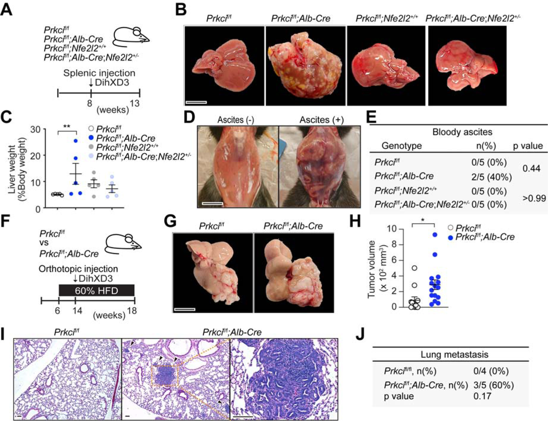
Figure 6. PKCλ/ι Deficiency in Non-tumorous Liver Tissue Generates a Pro-tumorigenic Microenvironment.

(A) Schematic representation of splenic injection of DihXD3.
(B-E) Images of transplanted livers (B), quantification of liver weight normalized to body weight (n = 5)
(C) and images (D), and incidence of bloody ascites (E) of mice treated as in (A).
(F) Schematic of orthotopic implantation of DihXD3 cells into livers of Prkcif/f and Prkcif/f;Alb-Cre male mice.
(G-J) Liver images (G), hepatic tumor volume (Prkcif/f, n = 12; Prkcif/f;Alb-Cre, n = 15 tumors) (H), H&E staining (I) and incidence (J) of lung metastasis (Prkcif/f, n = 4; Prkcif/f;Alb-Cre, n = 5 animals) of mice treated as in (F).
Mean ± SEM. *p < 0.05, **p < 0.01. Scale bar, 1 cm (B and G); 100 μm (I).
