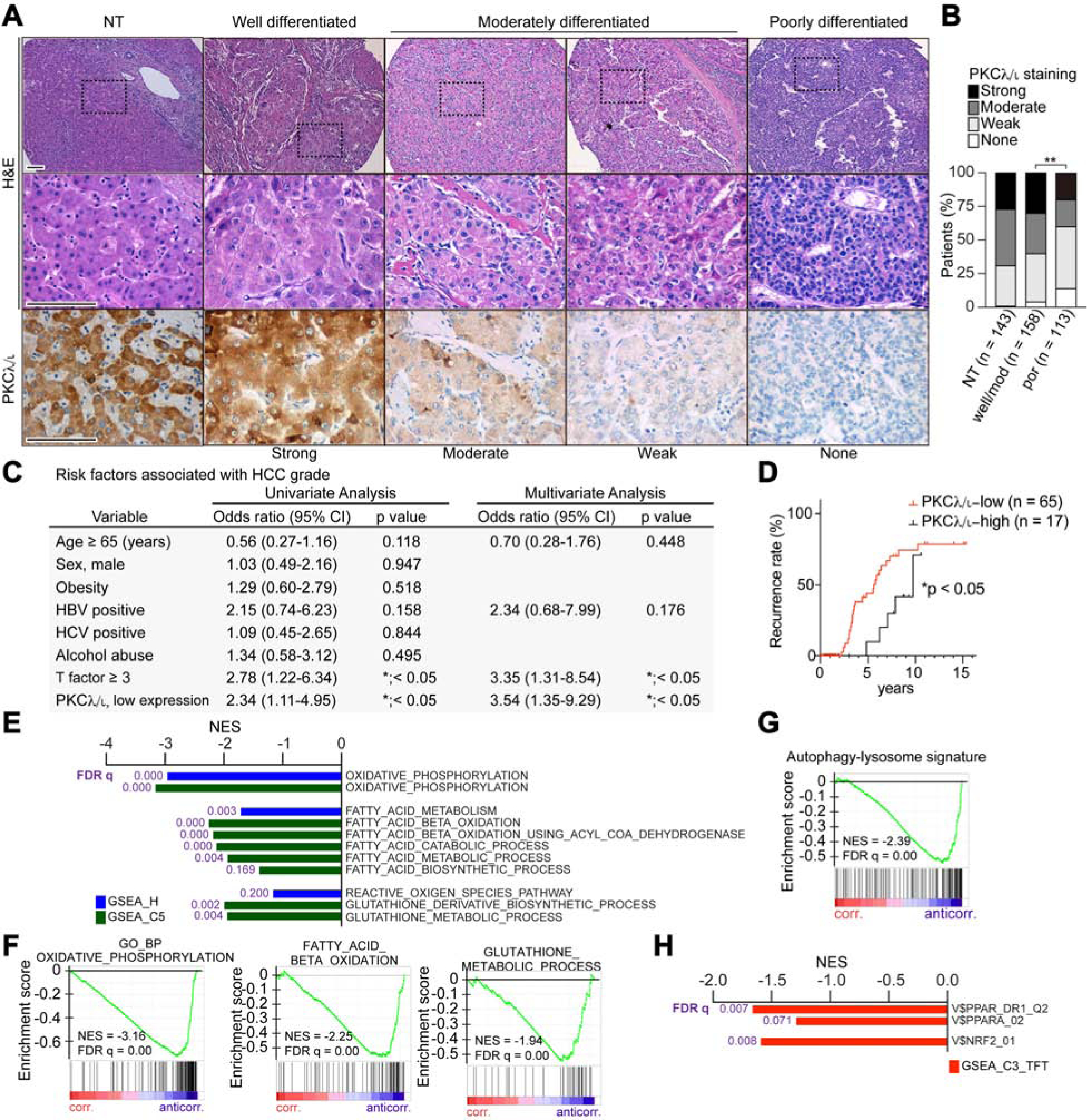
Figure 7. Low PKCλ/ι Expression in Human Liver Tissue is a Risk Factor for Late HCC Recurrence.

(A) H&E staining and IHC for PKCλ/ι in clinical HCC sections of different histological grade (n = 271 cases) and surrounding non-tumorous (NT, n = 143 cases) liver tissues. Scale bar, 100 μm.
(B) PKCλ/ι expression levels in HCC according to the IHC based classification; none, weak, moderate and strong (NT, n = 143; HCC, n = 271).
(C) Univariate and multivariate analyses to determine risk factors associated with HCC grade (odds ratio; poorly differentiated HCC vs HCC of well or moderately differentiated HCC), (n = 143).
(D) Kaplan-Meier curves of time to late recurrence in patients who underwent surgical resection of HCC. Patients were classified according to the PKCλ/ι expression level in surrounding non-tumorous liver tissues (n = 82).
(E) Negatively correlated pathways to Prkci gene expression in background liver tissues of HCC patients from TCGA dataset using compilation H and C5 (MSigDb).
(F-G) GSEA of the indicated genesets.
(H) Negative correlation between Prkci gene expression in background liver tissues of HCC patients from TCGA dataset with PPARα and NRF2 targets (C3, MSigDb).
*p < 0.05, **p < 0.01.
