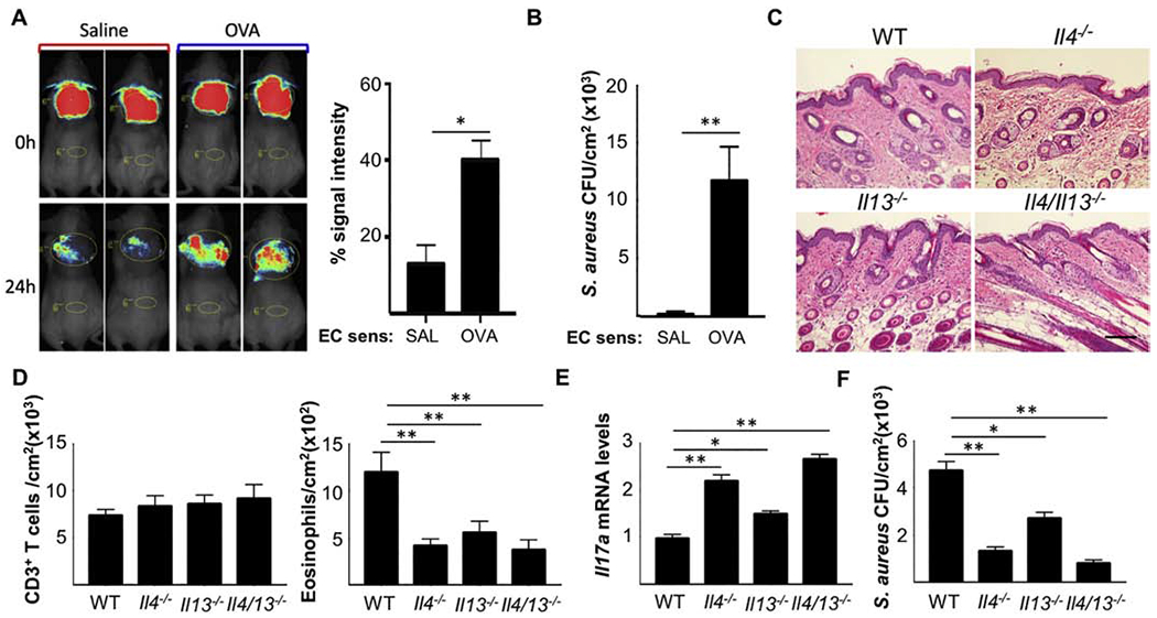
Figure 2. IL-4 and IL-13 impair S. aureus clearance from sites of allergic skin inflammation.

A. Representative in vivo imaging (left) and quantitation (right) of S. aureus fluorescence. B. S. aureus CFUs in skin homogenates. C. Representative H&E sections. Scale bar: 100 mm. D. Numbers of CD3+ T cells (left) and eosinophils (right) in skin. E. Skin Il17a mRNA levels F. S. aureus CFUs in the OVA sensitized skin of Il4−/−, Il13−/− and Il4/13−/− mice and WT controls. Results in B-F are representative of 2 independent experiments with 4-5 mice/group. Bars represent means±SEM. * = p<0.05, ** =p<0.005. Significance was calculated by using two-tailed Student’s t test.
