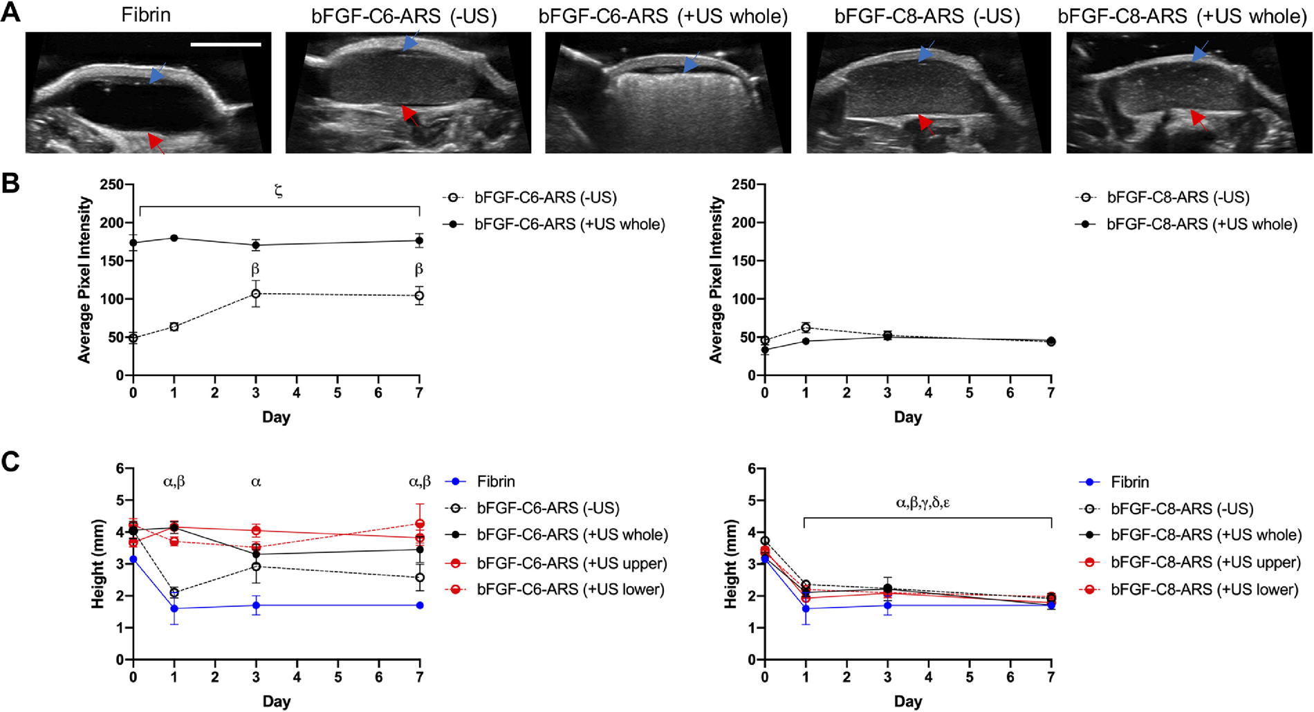
Figure 2.

ARSs were polymerized and exposed ex situ to US to generate acoustic droplet vaporization (ADV). The ARSs were implanted subcutaneously and longitudinally monitored in vivo using B-mode US. A) Transverse, B-mode images at 25 MHz show the in situ morphologies of the subcutaneously-implanted scaffolds on day 0. The blue arrow denotes the top interface of the ARS which was proximal to the overlying skin. The red arrow denotes the bottom interface of the ARS which was proximal to the underlying skeletal muscle. Scale bar for all images: 5 mm. The echogenicities (B) and heights (C) of ARSs containing bFGF-C6-emulsion (left) and bFGF-C8-emulsion (right) are displayed. Data are represented as mean ± standard error of the mean (N=5 per condition). Statistically significant differences (p < 0.05) are denoted as follows: α: vs. fibrin (day 0), β: vs. ARS (−US) (day 0), γ: vs ARS (+US whole) (day 0), δ: vs ARS (+US upper) (day 0), ε: vs ARS (+US lower) (day 0), and ζ vs. vs bFGF-C6-ARS (−US).
