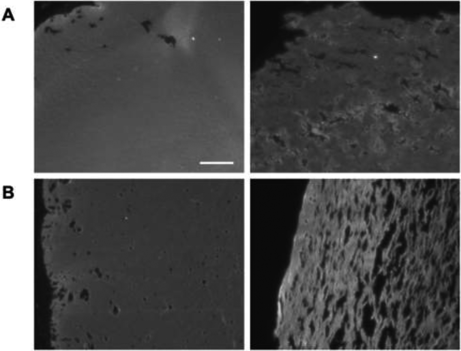
Figure 4.

Fluorescence images of A) fibrin (only) scaffolds and B) C8-ARSs highlight the increase in macroporosity with US exposure (right column) compared to −US condition (left column). Scaffolds, which contained Alexa Fluor 647-labeled fibrinogen, were polymerized and exposed to US ex situ prior to subcutaneous implantation. After 7 days, the scaffolds and surrounding tissue were harvested. Scale bar for all images: 100 μm
