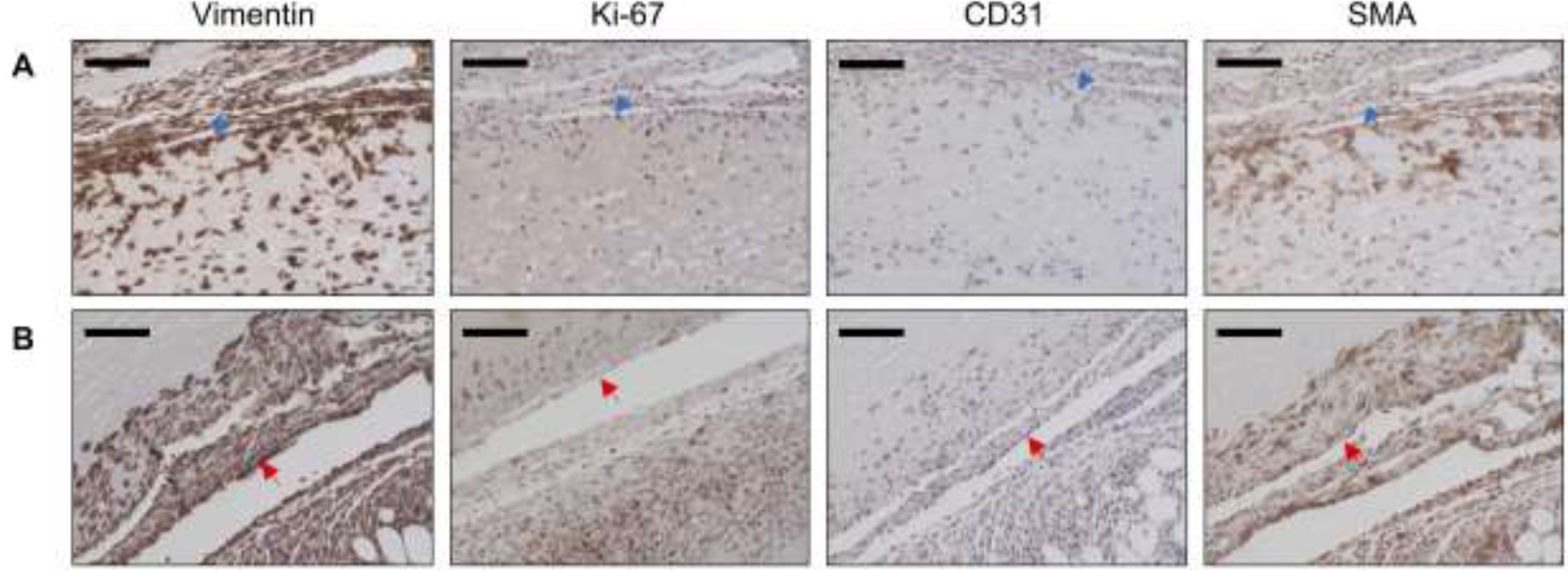
Figure 7.

Explanted ARSs were immunohistochemically stained for vimentin, Ki-67, CD31, and SMA. Panels A and B show upper and lower regions of bFGF-C8-ARSs exposed to the +US upper and +US lower patterns, respectively. The blue arrow denotes the top interface of the ARS which was proximal to the overlying skin. The red arrow denotes the bottom interface of the ARS which was proximal to the underlying skeletal muscle. Sections were counterstained with hematoxylin. Scale bar for all images: 100 μm.
