
Figure 1. μCOP printing of a 3D cardiac tissue.
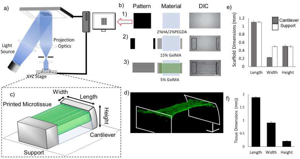
a) Schematic of the μCOP printing system. b) Mask-set and print order of: 1) 2%HAGM/2% PEGDA, 2) 15% GelMA, 3) 5% GelMA/NMVCM with DIC images (scale bar 250 μm). c) 3D schematic of full 3D tissue-measuring scaffold. d) Confocal 3D reconstruction of NMVCM cardiac tissue stained for actin (red) and nuclei (blue) across pillars. e) Measured scaffold dimensions in mm (n=19) f) Initial tissue dimensions of the 3D-printed scaffolds in mm (n=6)