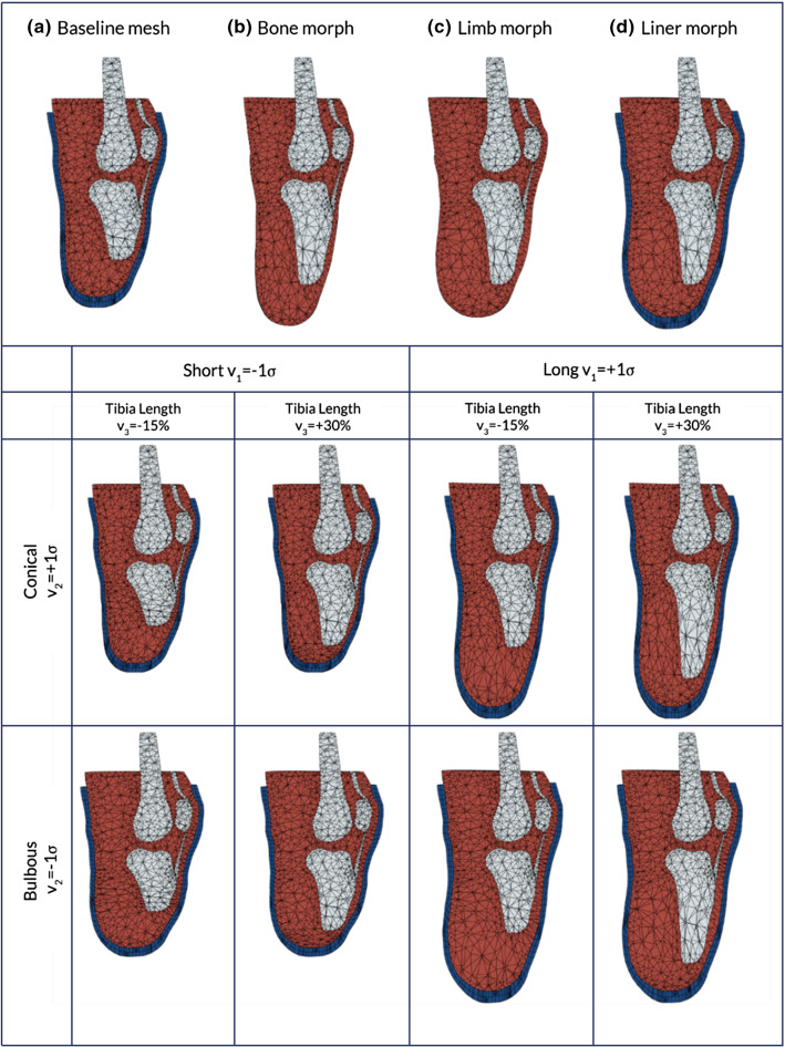
Fig. 3.
Procedure used for morphing the baseline FE mesh (a), through modifying the bone length (b), then morphing the external shape of the limb to match the SSM (c) and finally morphing the external liner (d). The result of morphing the FE mesh to morphological parameters of the model informed by residuum length, v1, residuum profile, v2 and tibia length, v3. Soft tissue is displayed as red, bone as grey and the liner as blue. The mesh has been visualised using a planar cut that goes through the elements

