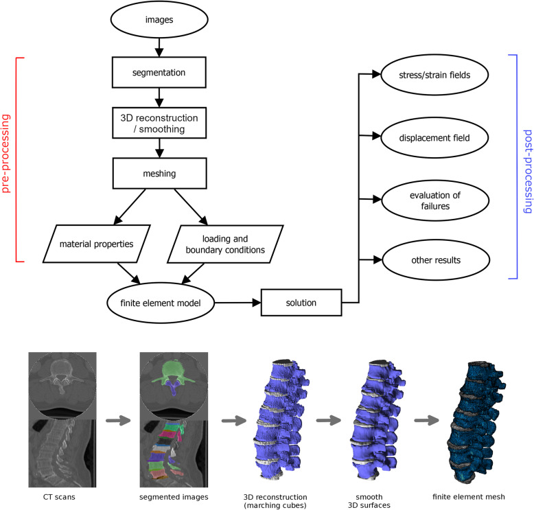
Fig. 1.
The workflow for the development and use of a finite element model from medical images (for example computed tomography scans): segmentation, three-dimensional reconstruction, improvement of the quality of the reconstructed surfaces by means of filtering (smoothing), meshing, assignment of loading/boundary conditions, and of material properties. The model can then be used to make predictions about stresses, strains, displacements, evaluating the failure of the materials, etc. Partially reprinted with permission from [1]

