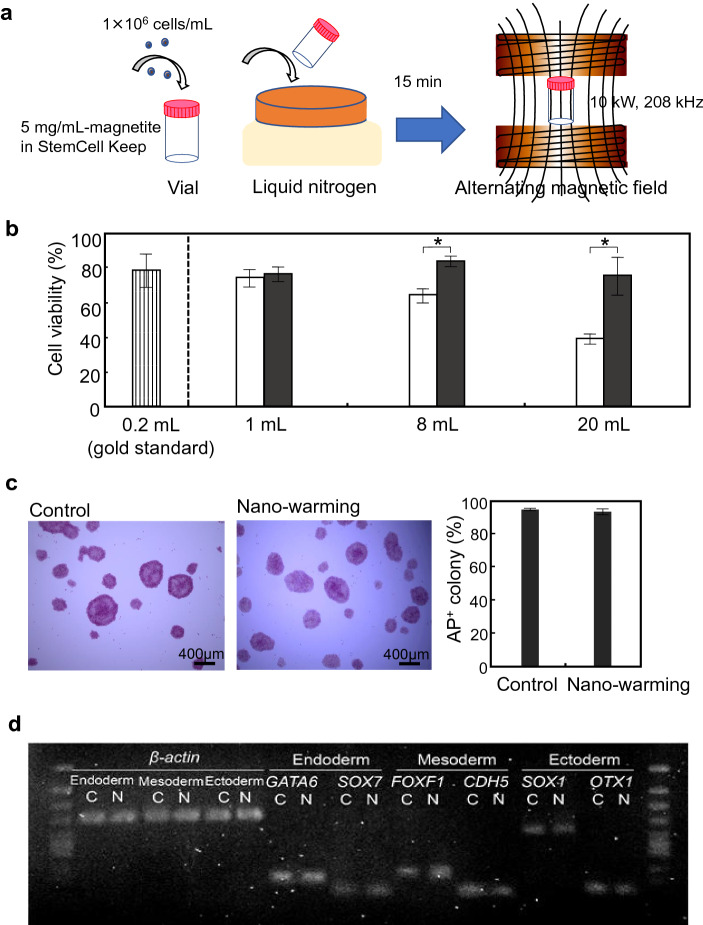
Figure 4.
Effects of nano-warming on hiPSC cryopreservation. (a) Schematic illustration for cryopreservation of iPSC cells. hiPSCs were applied to a vial at 1 × 10⁶ cells/mL in StemCell Keep containing magnetite nanoparticles at 5 mg/mL. The vial was directly immersed in liquid nitrogen for 15 min. The vial was then irradiated with an alternating magnetic field at 10 kW, 208 kHz. (b) Effects of nano-warming on hiPSC viability. After freezing and thawing, cell viability was assayed using a cell viability imaging kit based on Hoechst 33342 for live cells and SYTOX green nucleic acid stain for dead cells. Open columns, convective warming (water bath at 37 °C) of magnetite-loaded StemCell Keep; Closed columns, nano-warming of magnetite-loaded StemCell Keep. Data are expressed as the mean ± SD (n = 3). *P < 0.05. (c) Alkaline phosphatase (AP) staining of hiPSC colonies after nano-warming. Control, non-frozen hiPSCs. Data are expressed as the mean ± SD (n = 3). (d) RT-PCR analyses of lineage-specific genes. After nano-warming, hiPSCs were cultured under each differentiation condition for the three germ layers: endoderm, mesoderm, and ectoderm. C non-frozen control, N nano-warming. Relative expression levels normalized to β-actin gene expression are shown below the bands.

