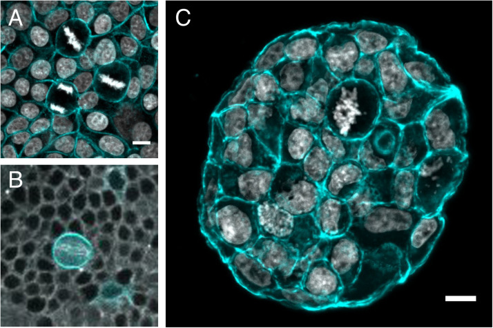
FIGURE 3.
Mitotic rounding in tissues and tumoroids. Examples of mitotic cells rounding while surrounded by other cells (A) in a non-transformed confluent epithelial cell monolayer (MCF10A) plated on a soft polyacrylamide hydrogel, stained with phalloidin-TRITC to visualize actin (cyan) and DAPI to visualize DNA (Gray) (Image by HM), (B) in vivo in a mitotic sensory organ precursor cell (labeled with LifeAct-GFP in cyan) in the notum of the developing Drosophila pupa. The whole tissue is labeled with tubulin (gray) to stain the mitotic spindle. (Image by Nelio Rodrigues) and (C) frozen section of an MCF-7 tumor spheroid grown for 14 days within a PEG/heparin hydrogel in 3D, stained with phalloidin-TRITC (cyan)/DAPI(gray) for F-actin/nuclei (image by AT). Scale bars are 10 μm.

