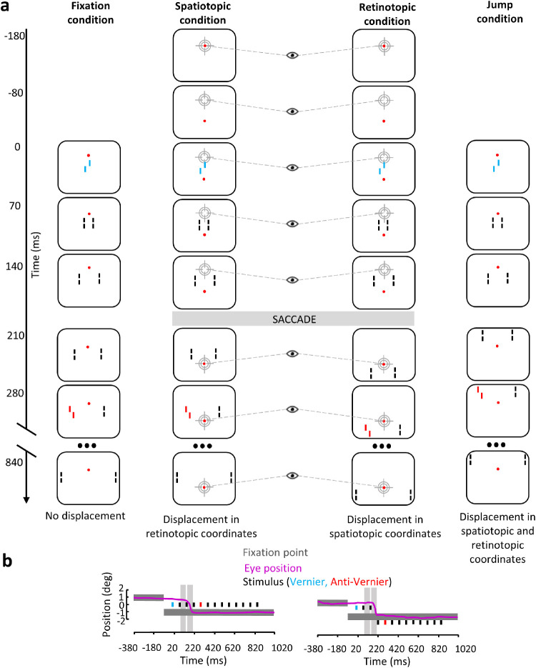
Figure 2.
(a) In the fixation condition, participants fixated the fixation point displayed 1° above the center of the screen during the entire trial. The SQM was presented in the center of screen. Here is shown the V-AV condition in which the central line and the fourth flanking line have equal contribution and are offset in opposite directions. In the spatiotopic condition, observers were instructed to make a saccade to the fixation point as soon as it was displaced. The SQM was displayed in the center of the screen; thus, there was no displacement in spatiotopic coordinates compared to the fixation condition, but there was retinal displacement. In the retinotopic condition, observers were instructed to make a saccade to the fixation point as soon as it was displaced. The SQM was first presented in the center of the screen and then displayed 1° below the new location of the fixation point after the saccade was detected; thus, the retinal stimulation was the same as in the fixation condition, but there was displacement in spatiotopic coordinates. In the jump condition, observers fixated the fixation point displayed 1° above the center of the screen during the entire trial. The SQM was first presented in the center of the screen. At 210 ms SOA, the sequence continued 1° above the fixation point; thus, the SQM was displaced in both spatiotopic and retinotopic coordinates compared to the fixation condition. Only the vernier–anti-vernier configuration is shown. (b) Time course of events with a sample eye position trace (pink line) in the spatiotopic and retinotopic conditions. Black rectangles indicate presentation of the individual lines of the SQM. The vernier and anti-vernier offsets are represented in blue and red, respectively. Dark gray rectangles indicate presentation of the fixation point. As described in the Methods section, only saccades made during the blank periods (light gray areas) were kept for analysis; that is, no saccade-induced motion smear occurred. Colors are for illustration purposes only; stimuli were white on a black background.

