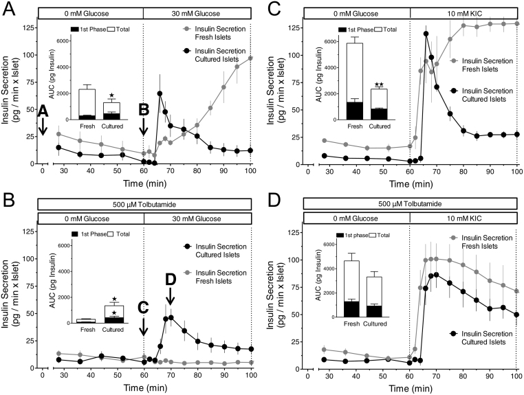
Figure 1.
Predominance of the first phase response in cultured mouse islets (black symbols) as compared to the variable response pattern in freshly isolated mouse islets (grey symbols). (A) 22-h-cultured islets were perifused with Krebs–Ringer medium containing 0 mM glucose for 60 min, then 30 mM glucose was present for 40 min. The increase of the glucose concentration from 0 to 30 mM glucose elicited virtually opposite kinetics of secretion in fresh and in cultured islets. (B) Same protocol as in A with 500 µM tolbutamide additionally present throughout the perifusion. Note a virtually complete loss of glucose stimulation with fresh islets but not with cultured islets. (C) Same protocol as in A except for using 10 mM KIC instead of 30 mM glucose as the nutrient secretagogue. The KIC-induced high level of secretion was only maintained in fresh islets. (D) Same protocol as in B except for using 10 mM KIC instead of 30 mM glucose as the nutrient secretagogue. Note the robust response of fresh islets to KIC stimulation as compared to the loss of glucose stimulation in B. Values are means ± s.e.m. of 4–5 experiments each. The inset shows the integrated insulin secretion from 60 to 75 min (first phase) and from 60 to 100 min (total). *P < 0.05, **P < 0.01, cultured vs fresh islets under the same condition, unpaired two-sided t-test. The secretion data obtained with fresh islets are taken from Schulze et al. (16).

 This work is licensed under a