Fig. 2.
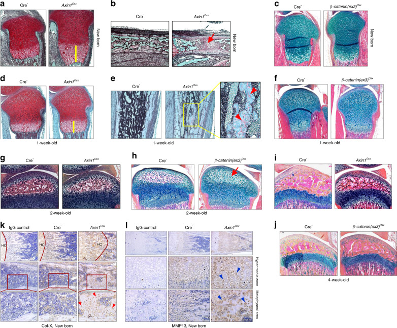
Delayed postnatal bone growth in Axin1Osx KO mice. a, d Results of Safranin O/Fast green staining revealed an expanded hypertrophic zone (yellow bars) in tibial growth plates of newborn and 1-week-old Axin1Osx KO mice compared with those of Cre-negative littermates. b Results of Alcian blue/Hematoxylin Orange G (AB/HO) staining showed that formation of a primary ossification center was delayed in newborn Axin1Osx KO mice (red arrow). e Results of Alcian blue staining showed that trabecular bone with large amounts of uncalcified osteoid (red arrowheads) was found in 1-week-old Axin1Osx KO mice. c, f No significant changes in the length and morphology of growth plate cartilage were seen in newborn and 1-week-old β-catenin(ex3)Osx activation mice compared with their Cre-negative littermates. g, h Results of Alcian blue staining showed a slightly delayed formation of a secondary ossification center in 2-week-old Axin1Osx KO mice. In contrast, a significantly delayed formation of a secondary ossification center (red arrow) was found in 2-week-old β-catenin(ex3)Osx activation mice. i, j Results of Alcian blue staining histology showed relatively normal hypertrophic cartilage development in 4-week-old Axin1Osx KO mice and in 4-week-old β-catenin(ex3)Osx activation mice. k, l Results of IHC showed that collagen type X (Col-X)-positive hypertrophic chondrocytes (HC) (red arrowheads) and MMP13-positive hypertrophic chondrocytes (blue arrowheads) were found in the metaphyseal bone area and in the middle of the bone marrow cavity of newborn Axin1Osx KO tibiae

