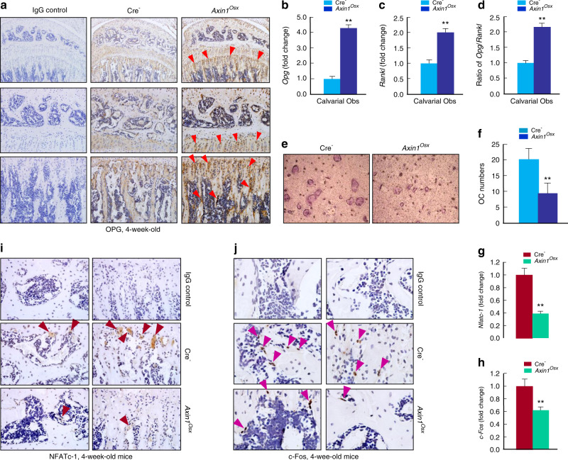
Fig. 5.
Alterations of OPG, NFATc-1, and c-Fos expression in Axin1Osx KO mice. a Results of IHC staining showed that more osteoprotegerin (OPG)-positive cells were observed in 4-week-old Axin1Osx KO mice, especially on the surface of trabecular bone (red arrowheads). b–d In calvarial osteoblasts isolated from newborn Axin1Osx KO mice, both Opg and Rankl expression was increased, and the ratio of Opg/Rankl was significantly higher in bone marrow stromal (BMS) cells derived from Axin1Osx mice than that of Cre-negative mice (n = 4, **P < 0.01). e WT BMM cells were cultured with the conditioned medium (CM) collected from cultured calvarial osteoblasts of 4-week-old Axin1Osx KO mice and Cre-negative littermates, respectively. Osteoclast formation in the cells cultured with CM from the Axin1Osx KO calvarial cells was significantly decreased compared with that in the cells cultured with CM collected from Cre-negative osteoblasts. f Quantification results showed decreased osteoclast numbers in the Axin1Osx KO group (n = 6, **P < 0.01). g, h The total RNA was extracted from tibia of 4-week-old Cre-negative and Axin1Osx KO mice and Nfatc-1 and c-Fos mRNA expression was detected by real-time PCR (n = 4, **P < 0.01). i, j In tibiae of 4-week-old Axin1Osx KO mice, NFATc-1-positive staining cells (brown arrowheads) and c-Fos-positive staining cells (purple arrowheads) were decreased compared with those of Cre-negative control mice

