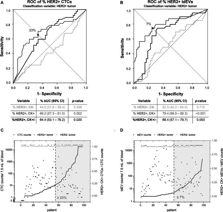
Fig. 4.
Prediction of HER2 status of tissue from CTCs and tdEVs. ROC curves of %HER2+CK− (light gray lines), HER2+CK+ (dark gray lines), and total HER2+CK+/− (black lines) CTCs (a) and tdEVs (b) treating HER2+ tissue (as assessed by FISH) as the classification variable. %HER2+CK+ populations performed the best as shown by the largest AUCs. The asterisks indicate the points, where sensitivity ≈ specificity for CTCs and tdEVs (23% HER2+CK+ CTCs leading to 65% sensitivity and 66% specificity; 7% HER2+CK+ tdEVs leading to 74% sensitivity and specificity). Scatter plot of total CTCs (c) and tdEVs (d) for each patient (x-axis). Samples were sorted on the percentage of HER2+CK+ CTCs or tdEVs respectively indicated by the dark gray lines. On the top of the panels and along the x-axis, the HER2 status of the tissue is indicated as positive (+) or negative (−), as evaluated by FISH. The vertical black dashed lines indicate the 23% HER2+ CTCs (and 7% HER2+ tdEVs) threshold, right of which the tissue of the patient could be considered as HER2+

