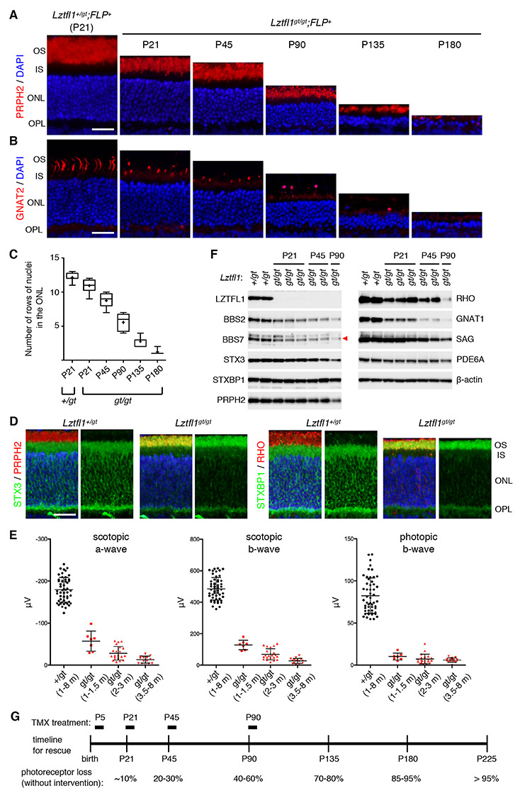
Figure 2.

Natural history of photoreceptor degeneration in Lztfl1gt/gt mice. (A) Progressive loss of photoreceptors in Lztfl1gt/gt mice. Retinal sections from Lztfl1+/gt;FLP+ and Lztfl1gt/gt;FLP+ mice at the indicated ages were stained with anti-PRPH2 antibodies (red) and DAPI (blue; for nuclei). OS, outer segment; IS, inner segment; ONL, outer nuclear layer; OPL, outer plexiform layer. Scale bar denotes 25 μm. (B) Loss of cones in Lztfl1gt/gt mice. Retinal sections were stained with anti-GNAT2 (red) antibodies. Others are the same as in (A). (C) Quantitation of the photoreceptor cell loss. Serial sections perpendicular to the retinal plane were collected from the central retina, and the number of rows of photoreceptor cell nuclei was counted (n = 4 per group; 3 sections per mouse; 2 locations per section (within 250 μm from the center)). Data are shown in a box-and-whisker plot. +, mean; error bars, minimum and maximum. (D) Mislocalization of STX3 and STXBP1 to the OS in Lztfl1gt/gt retinas at P13. Retinal sections from Lztfl1+/gt and Lztfl1gt/gt mice were decorated with anti-STX3 (green) and anti-STXBP1 (green) antibodies. Anti-PRPH2 (red) and anti-RHO (red) antibodies were used to delineate the OS. Merged images are shown on the left. DAPI (blue) was used to counterstain the nuclei. Scale bar denotes 25 μm. (E) Impaired photoresponse in Lztfl1gt/gt mice. Scotopic ERG a- and b-wave and photopic ERG b-wave amplitudes are shown in scatter plots. Each point represents the average value of two eyes from individual animals. Error bars represent standard deviation (SD) (n = 7–51). (F) Immunoblot analysis of whole eye protein extracts from Lztfl1+/gt and Lztfl1gt/gt mice. Eyes were collected at the indicated ages, and 50 μg of proteins weas loaded in each lane after normalization to total protein quantities. Each lane represents an individual animal. β-actin was used as a loading control. BBS7 band was marked with a red arrowhead. (G) Timeline of the rescue experiment. TMX was injected at postnatal (P) days P5/8/12 (P5 group), P21–25 (P21 group), P45–49 (P45 group) and P90–94 (P90 group) to restore Lztfl1 expression. Natural history of photoreceptor cell loss in Lztfl1gt/gt mice is summarized at the bottom.
