
Figure 2.
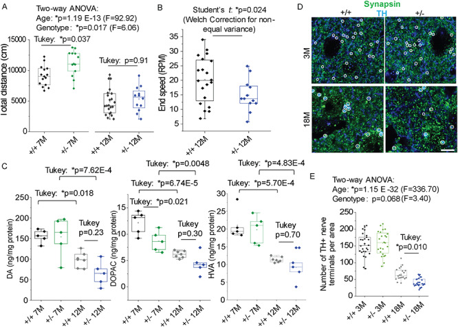
Synj1+/− mice display motor function abnormalities and progressive reduction of DAergic innervation. (A) Total distance measured in the open-field assay for male Synj1+/+ and Synj1+/− littermate mice at 7 months (7 M) and 12 months (12 M), respectively. 7 M cohort: Synj1+/+ (N = 15) and Synj1+/− (N = 13); 12 M cohort: Synj1+/+ (N = 22) and Synj1+/− (N = 13). Two-way ANOVA was performed, followed by Tukey’s post-hoc analyses. (B) Accelerated Rota-rod assay summarizing the end speed before mice fell off the rotating bar. P-values are from two-sample Student’s t tests with Welch’s correction. (C) Dopamine (DA) content and its metabolites levels in the striatum of 7-month-old (Synj1+/+ N = 5; Synj1+/− N = 5) and 12-month-old male mice (Synj1+/+ N = 7; Synj1+/− N = 5) measured by HPLC. Two-way ANOVA was performed, followed by Turkey’s post-hoc analyses. (D) Representative 3-month-old (3 M) and 18-month-old (18 M) striatal slices from a Synj1+/+ mouse and a Synj1+/− mouse immunolabeled with anti-TH and anti-synapsin1/2. Only puncta with dual immunofluorescence for TH (blue) and synapsin1/2 (green) were considered DAergic terminals (while circles) and were included in the double-blinded analysis. Scale bar, 5 μm. (E) Summary for the number of DAergic terminals analyzed from 3 male mice. DAergic terminals were sampled from 10–20 images from 2–3 brain slices for each individual mouse. Two-way ANOVA was performed, followed by Turkey’s post-hoc analyses.