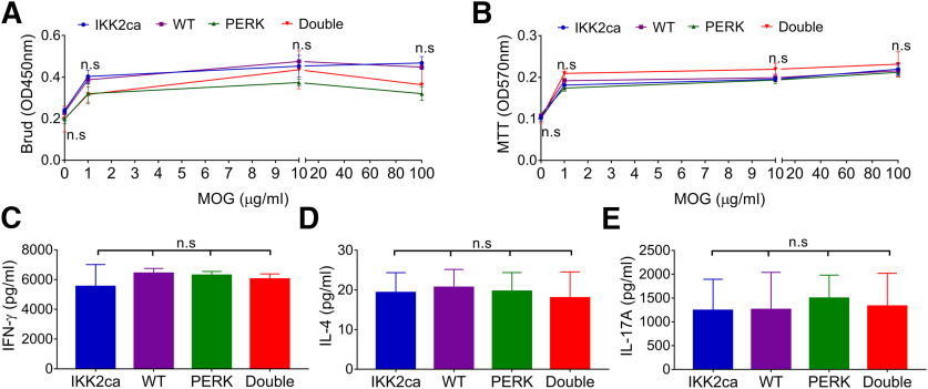
Figure 6.
Neither oligodendrocyte-specific expression of IKK2ca nor oligodendrocyte-specific PERK inactivation influenced T cell priming in the peripheral immune system during EAE. A, BrdU cell proliferation assay showed comparable T cell proliferation in IKK2ca cKI mice, WT mice, PERK cKO mice, and Double mice in response to MOG35-55 peptide. B, MTT cell viability assay showed comparable T cell viability in IKK2ca cKI mice, WT mice, PERK cKO mice, and Double mice in response to MOG35-55 peptide. C–E, ELISA analysis showed comparable ability of T cells to produce the cytokines IFN-γ, IL-4, or IL-17A in IKK2ca cKI mice, WT mice, PERK cKO mice, and Double mice in response to MOG35-55 peptide. N = 4 animals. Statistical analyses were done with a one-way ANOVA with a Tukey's post hoc test. Error bars represent SD; n.s., not significant.

