
Figure 4.
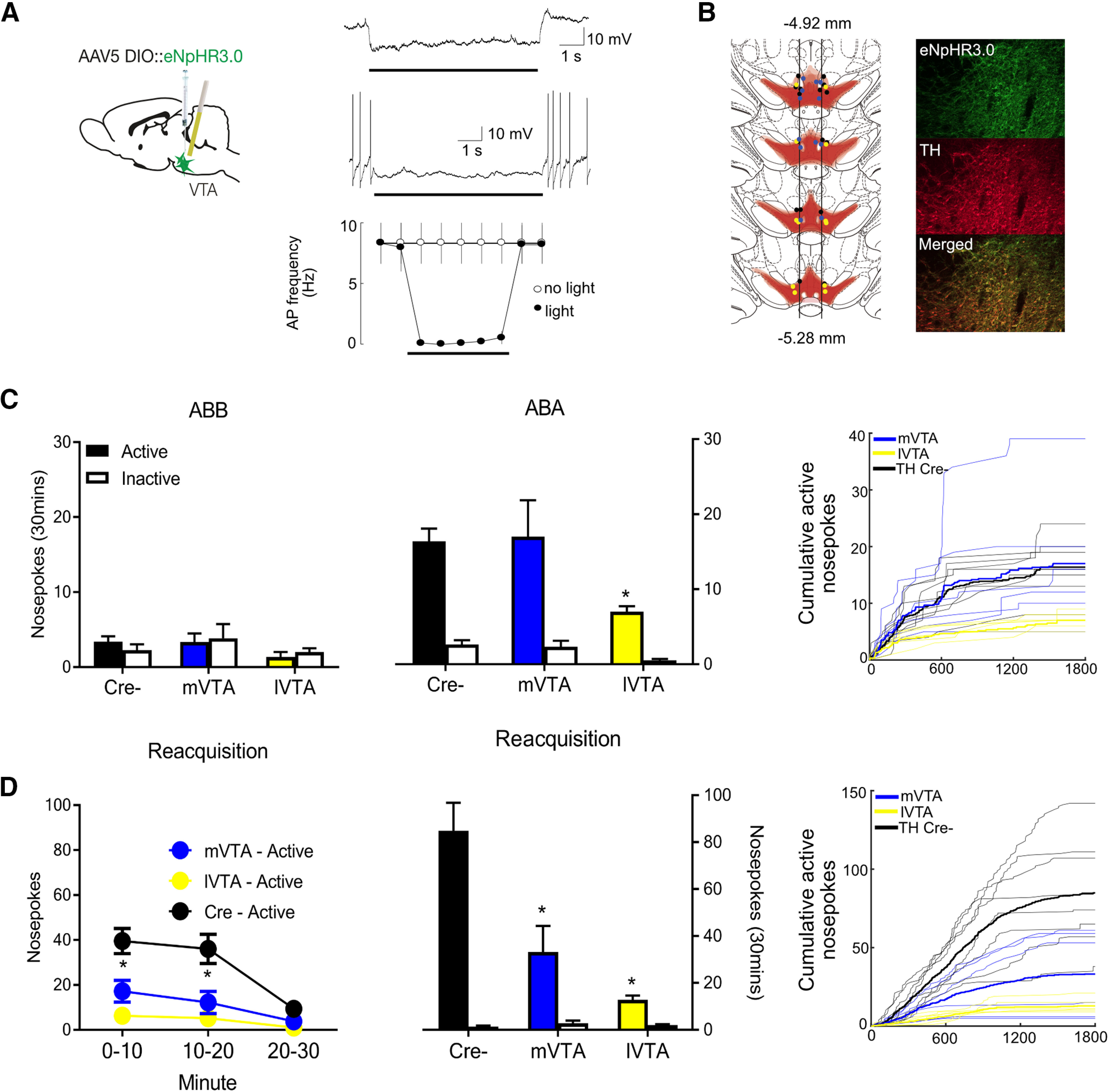
VTA TH optogenetic inhibition and relapse. A, Light-evoked responses of eNpHR3.0-positive VTA neurons. Typical light-evoked hyperpolarization, example of light-evoked suppression of spontaneous firing, and summary data plotting mean firing frequency evoked by a train of brief (5 ms) current injections in the presence and absence of light stimulation. Bars represent timing of light presentation. B, Location of eNpHR3.0 expression and fiber tips in midbrain with each rat represented at 25% opacity. The lateral edge of the fasciculus retroflexus was used as anatomic boundary between mVTA and lVTA. Example of eNpHR3.0 expression in VTA TH neurons. C, Mean ± SEM nosepokes during test in extinction and training contexts. Rats refrained from alcohol-seeking in the extinction context but relapsed to alcohol-seeking in the training context. lVTA, but not mVTA, inhibition prevented this relapse. Raw active nosepoke data are shown for all individual rats as cumulative functions. D, Mean ± SEM nosepokes during reacquisition. Both mVTA and lVTA inhibition reduced reacquisition. Raw active nosepoke data are shown for all individual rats as cumulative functions. Statistical analysis was done by ANOVA. *p < 0.05.