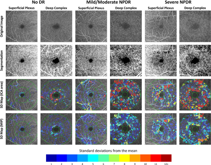
Figure 8.
Labeled standard deviation maps for subjects with no diabetic retinopathy, mild/moderate non-proliferative diabetic retinopathy, or severe non-proliferative diabetic retinopathy. Original images of the DVC have been brightened for clarity. All intercapillary areas are labeled based on the number of standard deviations its maximum ischemic point and area exceeded the reference mean.

