Fig. 1.
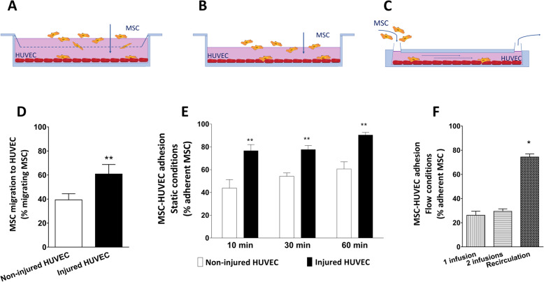
Migration and adhesion of MSC. a Migration of MSC is assessed by measuring the percentage of MSC able to migrate through a porous membrane towards (injured) HUVEC. b Adhesion of MSC to HUVEC in static conditions. MSC are added on a confluent monolayer of HUVEC, and the percentage of MSC which adhered after 10, 30, and 60 min is assessed by flow cytometry. c Adhesion of MSC to HUVEC in flow conditions. HUVEC are grown and injured in a flow chamber. MSC were infused 1 time or 2 times during flow or 1 time and recirculated for 10 min. The percentage of MSC which adhered was assessed by flow cytometry. d MSC showed an increased migratory capacity towards injured HUVEC compared to non-injured HUVEC. e MSC show increased adhesion to injured HUVEC compared to non-injured HUVEC. f MSC showed 28% adhesion capacity to injured HUVEC during flow conditions after one or two times infusion. Recirculation of MSC yielded increased adhesion of MSC to injured HUVEC during flow conditions. Significance of the comparison between 1 time infusion and recirculation is shown (n = 5). Results are shown as mean ± SD. **p value < 0.01; *p value < 0.05

