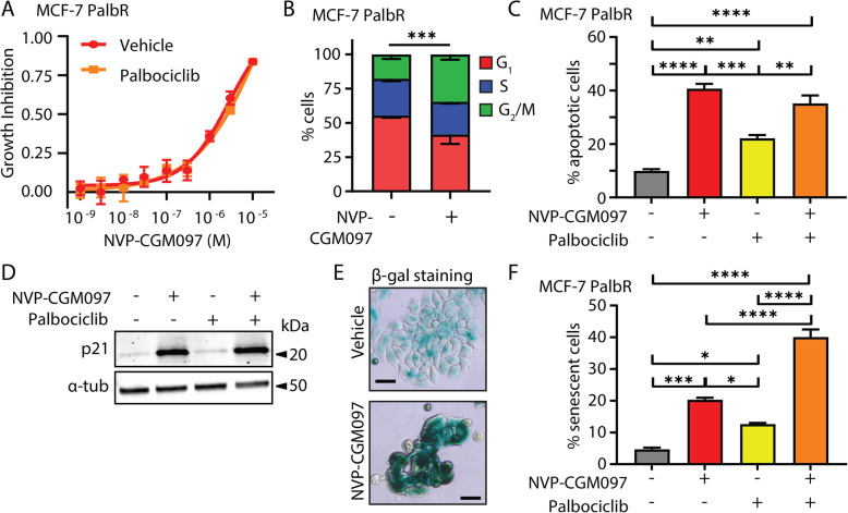
Fig. 7.
Palbociclib potentiates MDM2 inhibition via an increase in senescence in palbociclib-resistant MCF-7. a Growth inhibition of MCF-7 PalbR relative to vehicle (0.01% DMSO) of escalating doses of NVP-CGM097 in the presence (red) or absence (yellow) of 500 nM palbociclib—the dose used to generate the resistant cell line. No significant difference was observed after 48 h of treatment. b Proportion of cells in each cell cycle phase from flow cytometric quantification of propidium iodide staining of genomic DNA in MCF-7 PalbR cells after 48 h treatment with 1 μM NVP-CGM097 compared to vehicle (500 nM palbociclib, 0.01% DMSO). Statistical significance from χ2 test using the vehicle-treated profile as the expected value is indicated. Red = G1 (bottom), blue = S (middle), green = G2/M (top). c Proportion of MCF-7 PalbR cells staining positive for AnnexinV after treatment for 48 h with vehicle (0.01% DMSO, grey), 1 μM NVP-CGM097 (red), 500 nM palbociclib (yellow) or the combination of 1 μM NVP-CGM097 and 500 nM palbociclib (orange). Statistical significance from Tukey’s multiple comparison test is indicated above each column. d Western blot analysis of p21 in MCF-7 PalbR cells after 48 h incubation with 1 μM NVP-CGM097. α-tubulin staining is shown as an indication of relative loading. See Fig. S5D for full gel and blot images. e Representative images of MCF-7 PalbR cell cultures after exposure to vehicle (500 nM palbociclib, 0.01% DMSO) or 1 μM NVP-CGM097 for 48 h followed by staining for senescence-associated β-galactosidase. Bar = 20 μm. f Quantification of MCF-7 PalbR cells staining positive for senescence-associated β-galactosidase activity after treatment for 48 h with vehicle (0.01% DMSO, grey), 1 μM NVP-CGM097 (red), 500 nM palbociclib (yellow) or the combination of NVP-CGM097 and palbociclib (orange). Statistical significance from Tukey’s multiple comparison test is indicated above each column

