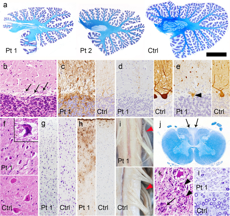
Fig. 1.
Neuropathologic findings. a Sagittal sections of the cerebellum showing diffuse atrophy of the folia and thinning of the dentate nucleus. The superior cerebellar peduncles are spared. Klüver-Barrera staining. b Loss of Purkinje cells and Bergmann gliosis (arrows) in the hemisphere. HE staining. c Bergmann gliosis is more evident by GFAP immunohistochemistry (IHC). d Decreased immunoreactivity of calbindin-D28k in the cerebellar cortex. The cell body and dendrites of a Purkinje cell are strongly stained in the control brain. Calbindin-D28k-IHC. e Retained parvalbumin immunoreactivity in the remaining basket and stellate cells in the cerebellar molecular layer, and an empty basket (arrowhead). The cell body and neurites of these interneurons in the cerebellar molecular layer, and those of a Purkinje cell are also stained in the control brain. Parvalbumin-IHC. f Although neurons in the dentate nucleus are shrunken (inset), their number is preserved. HE staining. g Moderate loss and shrinkage of neurons observed using Klüver-Barrera staining, and (h) gliosis detected by GFAP-IHC in the frontal cortex. i Atrophy of the cervical cord and posterior roots (arrowheads). j Atrophy and myelin pallor of the gracile fasciculus (arrows). Klüver-Barrera staining. k Loss of ganglion cells with a Nageotte nodule (arrow) and macrophage infiltration into the spaces where ganglion cells have been lost (arrowheads) in the dorsal root ganglion of the lumbar spinal cord. HE staining. (l) Severe loss of myelinated fibers in the sural nerve. Toluidine blue stain. Patient 1. Ctrl, control; Pt, patient. Bars = 1 cm in a; 8 mm in i; 300 μm in g, h, j; 100 μm in b-f, k; 50 μm in l.

