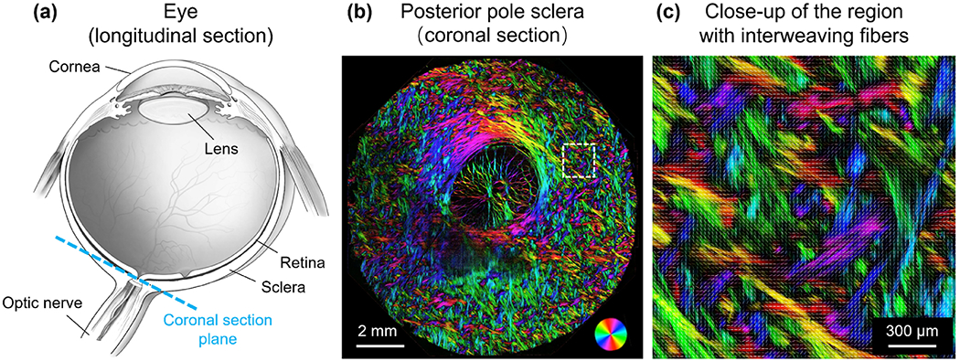
Figure 1.

(a) Schematic of an eye sectioned longitudinally. (b) Polarized light microscopy image of a coronal section of the posterior pole sclera in a sheep eye. Colors indicate local fiber orientation, whereas intensity is proportional to collagen fiber density. (c) Close-up of a region in the sclera exhibiting interweaving fibers.
