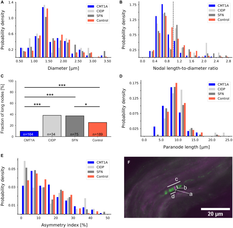
Figure 6.
Nodal and paranodal parameters. (A) Distributions of axonal diameter of CMT1A, CIDP, SFN and healthy control groups did not show any significant differences. (B) Distributions of ratios between the measured nodal gap length and the nodal diameter. Nodes with a ratio of >1 were considered long. The dashed line marks the ratio of 1. There were significantly fewer long nodes in the CMT1A group than in the other groups (CIDP: P < 0.005, SFN: P < 0.001, control: P < 0.00001). (C) Fractions of long nodes in the four groups; binomial test; *P < 0.05, ***P < 0.001. (D) Distribution of paranodal lengths. In patients with CMT1A, paranodal length was significantly decreased compared with the CIDP and control groups (CIDP: P < 0.01, SFN: P = 0.08, control: P < 0.001). (E) Distributions of asymmetry indices did not differ between the groups. (F) Representative micrograph of a node of Ranvier double-labelled with anti-pan-sodium channel (magenta) and anti-Caspr (green). Measurements were taken as indicated: a: nodal length, b: axonal diameter, c: paranodal length, d: length of hemiparanode.

