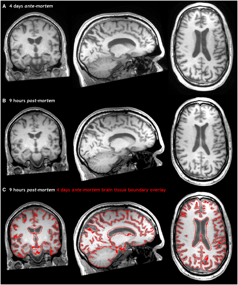
Figure 2.
Ante-mortem and post-mortem T1-weighted images of the brain. 3D T1-weighted images of the brain, 4 days ante-mortem (A) and 9 h post-mortem (B) registered in the same space. The fusion of both images (C) clearly shows an increase in brain volume post-mortem, particularly evident by smaller ventricles and the swollen cerebellum.

