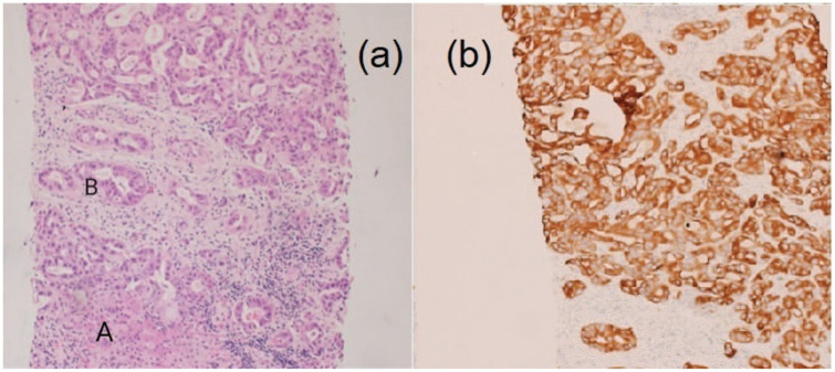
Figure 4.
(a) The photomicrographs show a normal liver tissue (A) exhibiting a tumor formed of cells arranged in glands and tubules, the tumor cells display hyperchromatic pleomorphic nuclei (B) with increased mitotic activity, the tumor is surrounded by desmoplastic stroma and (b) the tumor showed positive immunohistochemical reaction for CytoKeratin7 (CK7). The above-mentioned features are in keeping with cholangiocarcinoma.

