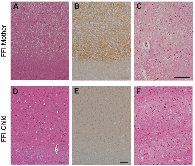
Figure 1.

Phenotypic features of FFI-Mother and FFI-Child. (A–F) Neuropathological features of FFI-mother and FFI-child. (A and D) HE stainings. The cerebral cortex showed widespread SC in FFI-Mother but not in FFI-Child. (B and E) Representative microscopic images of PrP immunostaining. The cerebral cortex showed widespread synaptic-type PrP deposition in FFI-Mother. No PrP deposition was observed in FFI-Child. (C and F) HE stainings. Neurons in inferior olive were preserved well in FFI-Mother, whereas inferior OD was observed in FFI-Child. Scale bars = 100 μm.
