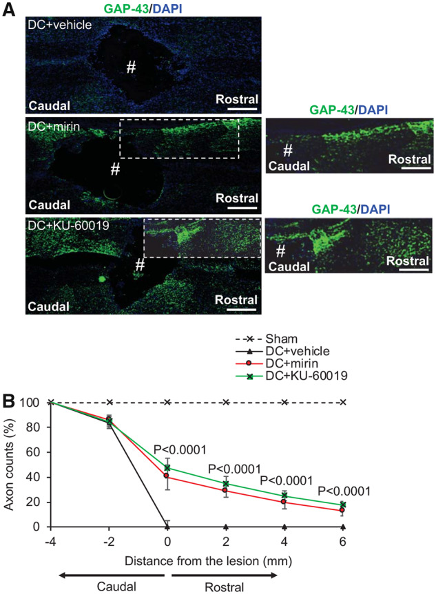
Figure 6.

Intrathecal injection of Mre11 and ATM inhibitors promote significant axon regeneration at 6 weeks after SCI. (A) GAP43+ (green) immunoreactivity was largely absent in DC + vehicle-treated groups (Blue = DAPI+ nuclei). However, in DC + mirin and DC+KU-60019-treated rats, enhanced GAP43+ immunoreactivity was present in the rostral segment of the spinal cord in beyond the lesion site (#) where a large cavity remained. (B) Quantification of the % of axons at different distances rostral to the lesion site showed significantly enhanced numbers of GAP43+ regenerating axons at all distances tested after treatment with DC + mirin and DC+KU-60019. (n = 10 nerves/condition). Scale bars in A = 200 µm. Comparisons in B by one-way ANOVA with Dunnet’s post hoc test (DC + vehicle versus DC+KU-60019).
