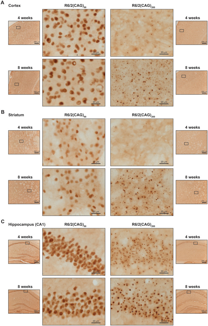
Figure 5.
Aggregated HTT accumulated in neuronal nuclei in the brains of R6/2(CAG)90 at a younger age than in R6/2(CAG)200 mice. The pattern of immunostaining detected with the MW8 antibody on brain sections from R6/2(CAG)90 and R6/2(CAG)200 mice at 4 and 8 weeks of age is shown for the (A) cortex, (B) striatum and (C) hippocampus. The location of the centre panel images in the brain sections is indicated in the adjacent thumbnails, and the position of the thumbnails within the tissue section is illustrated in Supplementary Figs 6 and 7. The wild type control sections are shown in Supplementary Figs 8 and 9. n = 3/R6/2 genotype and n = 1/wild type control. Centre panels, scale bar = 20 μm; thumbnails, scale bar = 200 μm.

