
Figure 7.
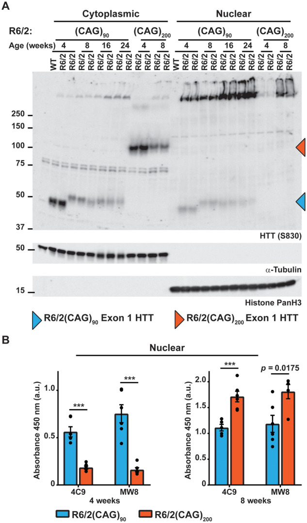
Soluble exon 1 HTT was detected in the nuclear fraction from R6/2(CAG)90 but not from R6/2(CAG)200 brains. (A) Western blot of cytoplasmic and nuclear fractions from R6/2(CAG)90 and R6/2(CAG)200 hemispheres at the ages indicated immunoprobed with the S830 antibody. A fragment that migrated as soluble exon 1 HTT was detected in the nuclear fraction of R6/2(CAG)90 but not R6/2(CAG)200 mice. S830 was used for the western blot presented here, as MW8 and 4C9 both detected cross-hybridising bands, which co-migrated with the R6/2(CAG)90 protein, making the presence of exon 1 HTT in the nuclear fraction harder to interpret. The purity of the nuclear and cytoplasmic fractions was indicated by immunoblotting with antibodies to α-tubulin and histone panH3 respectively. (B) Seprion ELISA analysis of HTT aggregation in nuclear fractions from R6/2(CAG)90 and R6/2(CAG)200 hemispheres at 4 and 8 weeks of age immunodetected with 4C9 or MW8 antibodies (n = 6/genotype). Statistical analysis was two-way ANOVA with Bonferroni post hoc correction. ***P < 0.001. The test statistic, degrees of freedom and P-values for the ANOVA are provided in Supplementary Table 6. WT = wild type.